At ImageWorks, we’ve been helping dental practices make the most of their dental imaging for decades. When it comes to dental cone beam systems, technology has come quite a way since we introduced our first generation 3D dental cone beam system in 2005 when we were called Dent-x.
As technology advances, we are always excited to introduce a dental professional to the power and value of 3D imaging, because it is compelling for so many offices. However, at the same time, with today’s 3D cone beam systems, we also like to say: with great power comes great responsibility. Because of this responsibility, we think it’s important to offer information that allows new users to enter the world of 3D with eyes wide open.
With this goal in mind, we thought it would be worthwhile to provide an overview of some of the most common questions that get asked when dental professionals are looking to implement a Cone Beam system in their office. We hope you find this useful.
What is the Field of View (FOV)?
The Field of View (many times referred to as FOV) refers to the size of the volume captured in the scan. Almost all CBCT systems on the market capture a volume that is shaped like a cylinder (i.e. a tin can). Therefore, the FOV is expressed with two numbers, which are typically in cm. The first number typically refers to the diameter of the circular face of the cylinder (the width of the tin can). The second number refers to the height.

The FOV typically advertised by a dental cone beam system will represent the largest FOV that can be captured by the system. However, almost all systems will offer the flexibility to perform smaller scans as well (in other words, a system that advertises a 16 x 8 FOV will also typically offer the option of scanning smaller volumes like 8 x 8, 8 x 10, or 4 x 6, etc. However, a system listing as a 8 x 8 FOV typically means that a larger scan is not an option with that system.
How big of an FOV do I need?
While there are many different options on the market, we sometimes put the different FOV sizes into three main groups.
First are the very large FOV machines are those that can capture most of the cranium in a single scan (e.g. largest scan size of 16 x 16 and above). These systems will be the most expensive and are typically used by oral surgeons or other specialists.
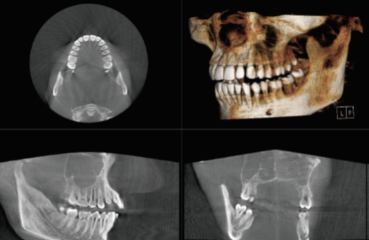
Second are the smaller FOV machines (e.g. 8 x 8 or 10 x 10). These can be an adequate dental cone beam system, as it allows capture of most of the essential anatomy. Below is a sample of a volume in this range (3D reconstruction, axial and sagittal views):

Potential challenges with cone beam systems that max out at this size may include:
- Cutting off 3rd molars
- Inability to perform airway analysis
- Inability to capture both TMJs in a single scan
- Little margin for error in patient positioning (i.e. suboptimal positioning may cause desired anatomy to fall outside the scanning volume)
Third are the medium size FOV dental cone beam machines (e.g. 16 x 8). Below are examples of a 16 x 8 volume (3D reconstruction, axial and sagittal views):

For most cone beam systems, the field of view represents the largest volume that can be captured. In other words, the user has the option to scan smaller volumes, but not larger.
In the end, the decision is a matter of what limitations the dental professional can tolerate versus the cost. Typically, the larger the field of view, the higher the price. However, at the same time, the larger field of view provides less limitations on what the user can capture.
Subscribe to Receive More Great Articles