For dental professionals that need both 3D volumes AND 2D panoramic radiographs to be high resolution.
Most dental cone beam systems use the same sensor for 2D panoramic radiographs as they use for the 3D scans. This means that one (or both) modality is typically suboptimal.
Here’s why this matters…
Learn more
See what our Customers are Saying


“We’ve been really happy to add the X-era to our practice, as it’s become such a crucial tool for me and my staff. We were excited to get a great price on a large field of view machine with this level of image quality. While I don’t use the 16 cm wide field of view all the time, it’s been critical for me to have this capability for certain cases. Wonderful product.” –Dr. Amr Hassan, Wesley Chapel, FL |
“I feel very strongly that it’s my job to make sure this practice is providing the best dental care that it can to our patients. Adding the cone beam to the office has provided us a new level of intelligence that enables me to advise my patients in ways I couldn’t fathom before. Traveling deeper into 3D dentistry has been a journey for all of us, and I’m very glad to have ImageWorks in my corner every step of the way.” –Dr Hieu Pham, Portland, OR |
“Our dentists are really impressed with the quality of the 3D images..” –Tim Baldwin, Woodbridge, VA |
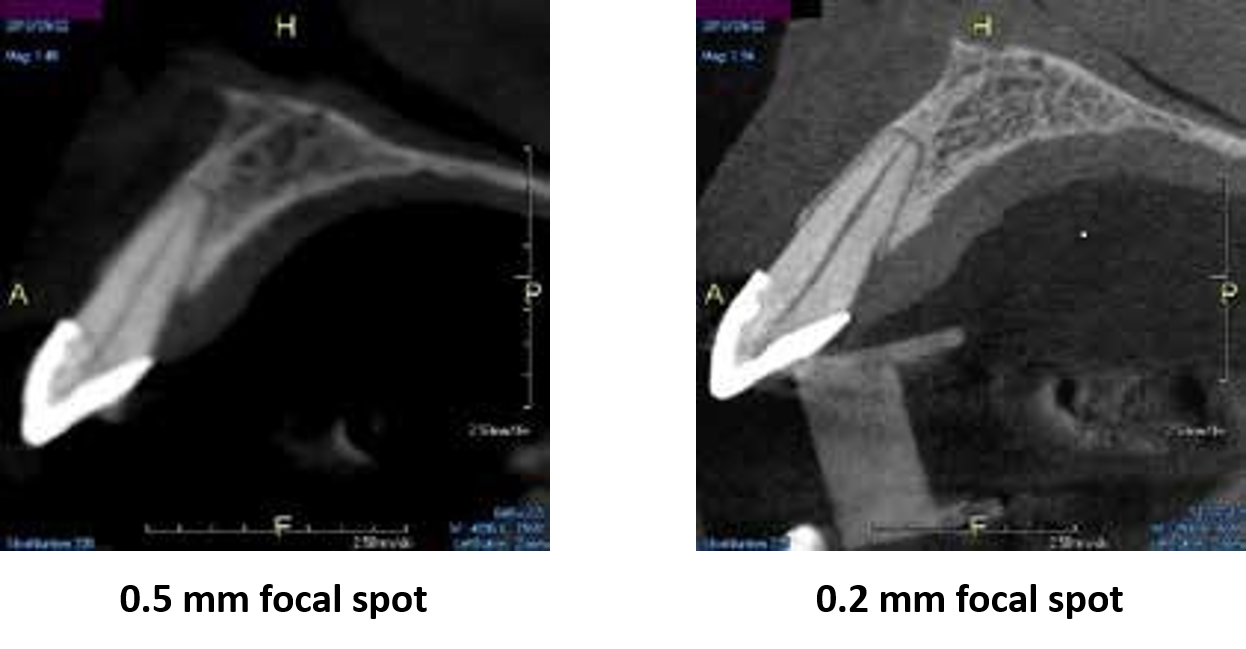
A 0.2 mm focal spot will allow you to spend less time convincing patients of your diagnosis and more time practicing dentistry.
Most cone beam systems either have a 0.5 mm or worse (larger focal spots equal less edge sharpness). The X-era focal spot is one of the smallest in the industry for a dental cone beam system. This not only means you and your staff can see more, but your patients can as well.
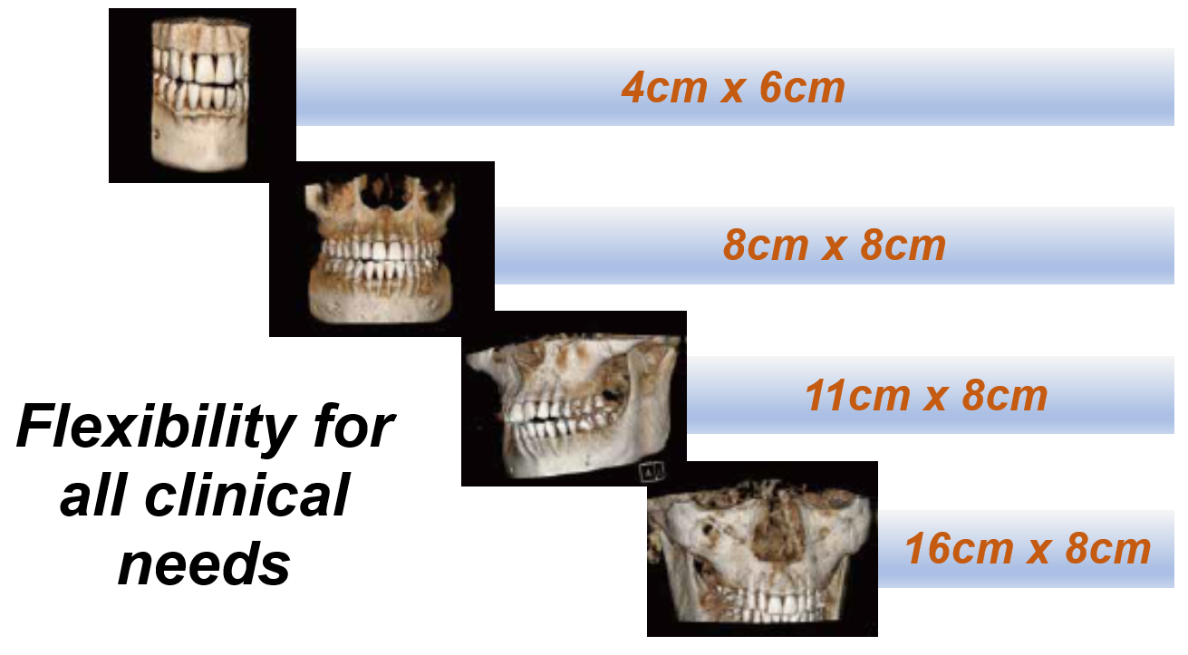
Field of View options to go as small as you want, or as big as you need.
Many dental cone beam systems max out at 9 or 10 cm wide. This may lead to:
- Cutting off 3rd molars
- Inability to perform airway analysis
- Inability to capture TMJs
- Little margin for error in patient positioning (i.e. suboptimal positioning may cause desired anatomy to fall outside the scanning volume which may require a retake)
The X-era gives the operator a number of options ranging from endo (4 cm x 6 cm) all way up to 16 cm wide. This offers fantastic flexibility and won’t limit what you can do with the platform.
Learn more
Low-dose options allow cone beam scans with lower radiation than traditional intraoral exams.
Traditional FMX studies can be 150 microsieverts or more of radiation for the patients. The low-dose modes of the X-era can provide a cone beam scan with less radiation while opening up a world of new information.
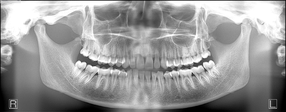
A Panoramic X-Ray to be proud of
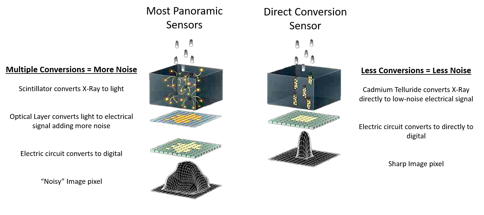
Most of our dental professionals depend on their cone beam system to produce a high-quality panoramic x-ray in addition to a high-quality 3D volume. Most dental cone beam systems sacrifice the performance of their panoramic modality by attempting to use the 3D sensor to perform 2D panos. The X-era uses a cone beam sensor for cone beam scans, and a panoramic sensor for panoramic scans.

In fact, the panoramic sensor is the same high-quality sensor that we use in our wildly successful pan-only platform, the Panoura 18S. Here’s what makes the panoramic modality so powerful…
What’s so great about a Direct Conversion Sensor?
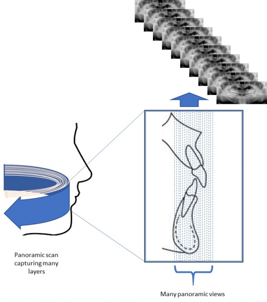
Every scan captures over 50 different panoramic layers!
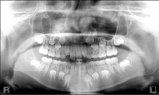
With most panoramic x-ray units, the arch of the patient needs to be exactly where the machine wants: along the path where the beam is focused.
- If positioning is not precise, a poor image can result.
- If the patient shifts slightly during the time the operator walks out to initiate the scan, a poor image can result.
- If the patient’s arch doesn’t match the trajectory of the scan, certain anatomy may not be in focus.
- The emergence profile of the anteriors could make it difficult to capture both mandibular and maxillary apices clearly in the panoramic scan.
The X-era also has Multi-Focal Plane capture capability. This means that every scan captures many layers both forward and backward. This creates a wide “envelope” where much more data is captured around where the patient is positioned.
Great images even if your staff’s technique is less than perfect!
Read Article: Why Can’t I see the Anterior Apices in My Panoramic?
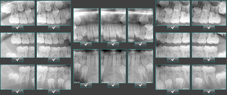
Not only Bitewings, but an entire FMX!
Vertical bitewing feature as well as FMX Clipping feature creates 18 intraoral-sized images clipped from the pan with one click.
Ideal tool for patients who won’t tolerate traditional PAs
- Instead of taking 18 individual images with an intraoral X-Ray, they can all be generated at once in 14 seconds.
Review Sample Images and Volumes
Support and Expertise to help you thrive!
We’ve deployed thousands of dental imaging solutions across over 30 years. Whether it’s a technology challenge, or a clinical challenge, our team has seen it all. You will have all that expertise on your team.
Integration is always a key question – because everything has to work well together. Our system is open source, so it is seamless with most other dental systems. In addition, our team has worked with almost every other software and hardware under the sun – so we can also help configure to your staff’s unique needs.
We focus on three things: Great Images, Great Pricing, and No Issues.
Learn more
Read Article: Two Very Different Approaches to 3D Dentistry
Why 3D?
The Secret Power to Get More Information From Your Panoramic
CBCT Case: Tooth fracture
CBCT Case: Patient with Sinus Trouble and how to work with ENTs
CBCT Case: Saving the tooth that was supporting a bridge
CBCT Case: Infection from sinus graft
CBCT Case: Identifying bone loss
CBCT Case: Drainage in Lower Left 2nd Molar
CBCT Case: Asymptomatic infection
FAQ
Will your system work with my software?
What training will be provided?
What happens if we need support?
Will your system work with my lab?
Can I share the volumes with colleagues who don’t have your software?
Do you offer financing?
If you would like to have
Enormous new diagnostic information unlocked and at your fingertips
All members of your staff fully confident using the tools so they can focus their attention on patients not devices
Full confidence that you have a diligent and experienced support team in your corner to help you succeed
Schedule a 15 minute call with one of our imaging specialists to see how we can assist.
Learn more